Gas chromatography–mass spectrometry

GCMS
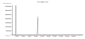
GC analysis is a common confirmation test. GC analysis separates all of the components in a sample and provides spectral output. The sample is injected in the GC port. The GC vaporizes the sample, separates and analyzes the various components. Each component produces a spectral peak that is recorded on a paper (intensity vs retention time). The time elapsed between injection and elution is called the “retention time.” The retention time differentiates the different components within the sample. The area under the peaks is proportional to the quantity of the corresponding substances in the sample analyzed. The peak is integrated to give the area under the peak. The bottom of the peak is the base line.
The different molecular weight compounds are eluded at different rates, lighter weight compounds elude first. The chemical nature may slow down the elution time.
The equipment consists of an injection port at one end of a metal column packed with substrate material and a detector at the other end of the column. A carrier gas propels the sample down the column. Flow meters and pressure gauges are used to maintain a constant gas flow. A gas that does not react with the sample or column is used, normally argon, helium, nitrogen, or hydrogen. Helium is used most often because it does not react. Hydrogen may react and convert the sample into another substance.
The sample enters the column in a discreet, compact packet. The injection port is maintained at a temperature at which the sample vaporizes immediately. The sample spreads evenly along the cross section of the column, forming a plug. The column is a metal tube, often packed with a sand-like material to promote maximum separation. Columns are pre-packed by vendors. As the gas moves through the column, the different molecular characteristics determine how each substance in the gas interacts with the column surface and packing. The column allows the various substances to partition themselves.
Substances that minimize surface interaction with the packing move through the column rapidly. Substances that adhere to the packing are impeded but eventually elute from the column. The various components should separate before eluting from the column end.
The GC instrument uses a detector to measure the different compounds as they emerge from the column. Among the available detectors are the argon ionization detector, flame ionization detector, flame emission detector, cross section detector, thermal conductivity detector, and the electron capture detector. Choosing the proper detector depends upon the use. Depending on further analysis a detector is chosen; the flame detectors destroy the sample, the thermal conductivity detector is universally sensitive, and the argon ionization detector requires argon as a carrier gas. The spectral output is stored electronically and displayed on a monitor.
The argon ionization detector does not detect water, carbon tetrachloride, nitrogen, oxygen, carbon dioxide, carbon monoxide, ethane, or compounds containing fluorine. The flame ionization detector does not respond to water, nitrogen, oxygen, carbon dioxide, carbon monoxide, helium, or argon. If a specimen contains water, a flame ionization detector should be used. The electron capture detector cannot detect simple hydrocarbons but does detect compounds containing halides, nitrogen, or phosphorus.
The amount of time that a compound is retained in the GC column is known as the retention time. Retention time is measured from the time the sample is injected until the time the compound elutes from the column. The retention time can aid in differentiating between some compounds. However, retention time is not a reliable factor to determine the identity of a compound. If two samples do not have equal retention times, those samples are not the same substance. However, identical retention times for two samples only indicate a possibility that the samples are the same substance.
Before analyzing a sample, the GC is tuned and calibrated. Tuning is accomplished using specific concentrations of Decafluorotriphenylphosphine and p-Bromofluorobenzene. A technician can process a spiked sample (containing a known concentration of a substance) to check calibration and tuning. If the GCMS instrument does not detect the substance or shows a greater or lesser concentration than the known concentration, the instrument must be re-calibrated. Also, a blank sample (containing no detectable compounds) can be used to test the GCMS instrument’s data reporting accuracy and performance. If the device indicates the presence of a substance in the blank sample, the device may contain residue from prior analysis. If this occurs, a re-tune and recalibration is needed.
Proper procedures require the spectral output with a known standard sample of the suspected substance. The standard sample must be analyzed with the same instrument, under the same conditions, immediately before and immediately after analyzing the unknown specimen. If the resulting three spectral outputs do not agree, then a reliable identification of the specimen based on the GC analysis is not possible.
Gaussian spectral peaks should be symmetrical, narrow, separate (not overlapping), and made with smooth lines. GC peaks that are broad, overlapping, or unevenly formed are unreliable. If a poorly shaped peak contains a steep front and a long, drawn-out tail, this may indicate traces of water in the specimen.
The sample should be injected into the septum rapidly and smoothly to attain good separation of the components in a sample. If the sample is injected too slowly, the peak may be broad or overlap. A twin peak may result from the technician hesitating during the injection. A smoothly performed injection, without abrupt changes, should result in a smoothly formed peak. A twin peak may also indicate the injection contained two samples concurrently.
The area under the peak is proportional to the amount of the substance that reaches the detector. No detector responds equally to different compounds. Results using one detector will probably differ from results obtained using another detector. Therefore, comparing analytical results to tabulated experimental data using a different detector does not provide a reliable identification of the specimen.
A “response factor” must be calculated for each substance with a particular detector. A response factor is obtained experimentally by analyzing a known quantity of the substance into the GC instrument and measuring the area of the relevant peak. The experimental conditions (temperature, pressure, carrier gas flow rate) must be identical to those used to analyze the specimen. The response factor equals the area of the spectral peak divided by the weight or volume of the substance injected.
The temperature of the GC injection port must be high enough to vaporize a liquid sample instantaneously. If the temperature is too low, separation is poor and broad spectral peaks should result or no peak develops at all. If the injection temperature is too high, the specimen may decompose or change its structure. If this occurs, the GC results will indicate the presence of compounds that were not in the original specimen.
If any substance remains inside the column, the substance may elute during subsequent analyses with other specimens. This may result in an unexpected peak in the output. The peak produced should be broad.
If the GC instrument uses hydrogen for the carrier gas, the question “does the hydrogen gas react with any of the sample components?” must be answered. If the hydrogen does react, a broad peak will result. When using a thermal conductivity detector, care should be taken as a false peak may occur if the carrier gas’s thermal conductivity is in the range of the thermal conductivity of any compound in the specimen. An unstable carrier gas flow rate may produce a drifting baseline and false broad peaks. A carrier gas should be 4 9s pure. Regular changing of the gas filter should prevent significant impurities.
GC analysis is highly reliable if the instrument is properly maintained, procedures followed, and the interpretation is competent. While some factors rarely affect GC analysis, some factors are absolutely essential for the use of reliable GC evidence. In all cases a technician must process a standard sample containing a verified composition identical to the presumed contents of the collected specimen. This standard sample must be processed before and after the collected specimen under identical conditions. Any output from the collected specimen that does not match the standard sample is inconclusive. If tabulated reference data exists for the relevant conditions, the specimen data must match the reference data.
Mass Spectrometry identifies substances by electrically charging the specimen molecules, accelerating them through a magnetic field, breaking the molecules into charged fragments and detecting the different charges. A spectral plot displays the mass of each fragment. One can use the compound’s mass spectrum for qualitative identification. The fragment masses are used to piece together the mass of the original molecule, the “parent mass.”
The parent mass is obtained by piecing together the fragment ions scattered though out the tic. From the molecular mass and the mass of the fragments, reference data is compared to determine the identity of the specimen. Each substance’s mass spectrum is unique. Providing that the interpretation of the output correctly determines the parent mass, MS identification is conclusive.
There are many different types of MS instruments; each one uses a different apparatus and process for producing mass spectra. This MS description will be a conventional large magnet mass spectrometer. The MS instrument contains a sample inlet, an ionization source, a molecule accelerator, and a detector.
MS analysis requires a pure gaseous sample. The sample inlet is maintained at a high temperature, up to 400° C (752° F), to ensure that the sample stays a gas. Next the specimen enters the ionization chamber. A beam of electrons is accelerated with a high voltage. The specimen molecules are shattered into well-defined fragments upon collision with the high voltage electrons. Each fragment is charged and travels to the accelerator as an individual particle.
In the acceleration chamber the charged particle’s velocity increases due to the influence of an accelerating voltage. For one value of voltage only one mass accelerates sufficiently to reach the detector. The accelerating voltage varies to cover a range of masses so that all fragments reach the detector.
The charged particles travel in a curved path towards the detector. When an individual charged particle collides with the detector surface, several electrons (also charged particles) emit from the detector surface. Next, these electrons accelerate towards a second surface, generating more electrons, which bombard another surface. Each electron carries a charge. Eventually, multiple collisions with multiple surfaces generate thousands of electrons which emit from the last surface. The result is an amplification of the original charge through a cascade of electrons arriving at the collector. At this point the instrument measures the charge and records the fragment mass as the mass is proportional to the detected charge.
The MS instrument produces the output by drawing an array of peaks on a chart, the “mass spectrum.” Each peak represents a value for a fragment mass. A peak’s height increases with the number of fragments detected with one particular mass. As in the case of the GC detectors, a peak may differ in height with the sensitivity of the detector used.
Each substance has a characteristic mass spectrum under particular controlled conditions. The sample can be identified by comparing the sample’s mass spectrum with known compounds. Quantitative analysis is possible by measuring the relative intensities of the mass spectra.
Usually a mass spectrum will display a peak for the unfragmented molecule of the specimen. This is commonly the greatest mass detected, called the “parent mass.” The parent mass reveals the mass of the molecule while the other peaks indicate the molecule’s structure.
Determining the parent peak and consequently the molecular mass of the specimen is the most difficult part of MS analysis.
The “resolution” is a value that represents the instrument’s ability to distinguish two particles of different masses. The greater the MS’s resolution, the greater its usefulness for analysis. An MS instrument provides more accurate results for larger molecules when the instrument has a high resolution. A high resolution MS instrument is advisable for analyzing body fluids because they have high molecular masses. A low resolution MS instrument may not sufficiently characterize a large mass substance.
If the interior pressure in an MS instrument is too high, erroneous results may occur. As the sample molecule breaks up, the fragments accelerate. If a fragment collides with another fragment, then these two fragments may combine to make a new particle. In this event, the detector will register the mass of this new particle on the mass spectrum. The reference spectra for comparison are produced under low pressure conditions which minimize collisions between fragments. This minimizes the chance of finding a spectral peak where one is not expected.
Finding the correct parent peak in the mass spectra may be difficult. Finding the parent peak helps to determine the parent mass, this should lead to determining the sample’s molecular mass.
High speed scanning MS instruments are able to rapidly analyze specimens. However, the increased speed is a tradeoff for decreased resolution. Quantitative measurements are unreliable with high speed scanning.
MS analysis is highly reliable if the instrument is of sufficient resolution and the interpretation of the results is competent. While some factors rarely affect MS analysis, some factors are absolutely essential for the use of reliable MS evidence. In all cases one must process a standard sample containing a verified composition identical to the presumed contents of the collected specimen. This standard sample must be processed under identical conditions, both before and after processing the collected specimen.
GCMS
The GC device is generally a reliable analytical instrument. The GC instrument is effective in separating compounds into their various components. However, the GC instrument cannot be used for reliable identification of specific substances. The MS instrument provides specific results but produces uncertain qualitative results. When an analyst uses the GC instrument to separate compounds before analysis with an MS instrument, a complementary relationship exists. The technician has access to both the retention times and mass spectral data.
GCMS analysis, where the effluent to the GC instrument is the feed to the MS instrument, is in wide use for confirmation testing of substances.
The GCMS has been used in:
- Arson investigations
- Engine exhaust analysis
- Petroleum product analysis
- Blood monitoring in surgery
- Drug testing
- Environmental contaminates
- Liquid identification and separation
- Deformulation- Lithium Ion Batteries